Which Muscle Forms The Floor Of The Mouth

Floor Of Mouth The Term Floor Of The Mouth Is Used Differently By Different Authors But In All Cases It I In 2020 Muscle Anatomy Facial Nerve Foot Reflexology Massage

Muscle Identification Anatomy Of The Neck Medical Anatomy Muscle Anatomy

The Geniohyoid Muscle Is A Narrow Muscle Situated Superior To The Medial Border Of The Mylohyoid Mus Dental Anatomy Human Anatomy And Physiology Muscle Anatomy

Mylohyoid Origin Insertion Action Nerve Supply Muscle Anatomy Facial Anatomy Human Body Anatomy

Esophagus Anatomy بحث Google Human Body Anatomy Human Anatomy

Tongue The Tongue Is An Extremely Mobile Mass Of Striated Muscle Covered By Mucous Membrane Ari In 2020 Facial Nerve Facial Nerve Branches Digestive System Anatomy

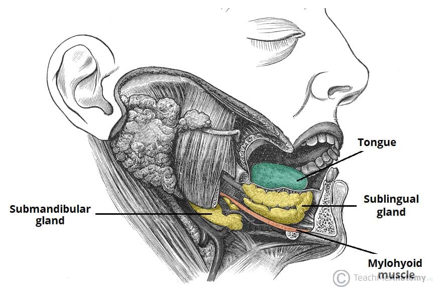
The squamous epithelium at the mucosal surface and other superficial anatomic components of the floor of the mouth are easily evaluated at physical examination the mylohyoid muscle sling is formed by the paired mylohyoid muscles which arise from the inner surface of the mandible extend from the symphysis anteriorly to the last molar tooth posteriorly and insert onto the posterior aspect.
Which muscle forms the floor of the mouth.

Floor Of The Mouth
Salivary Glands Numerous Glands Secrete The Watery Somewhat Viscous Fluid Known As Saliva Into The Oral Cavity Small Sal In 2020 Salivary Gland Facial Nerve Glands

Ranulas Ranulae And Their Treatment Salivary Gland Infection Salivary Gland Oral Pathology
The Oral Cavity Divisions Innervation Teachmeanatomy

Nasal Cavity Definition From Answers Com Nasal Cavity Paranasal Sinuses Nose Diagram

Pin By Lisa Redfern On Public Domain Misc Free Imagery Metallic Taste In Mouth Bad Breath Remedy Bad Breath

Septum Of The Genioglossus Muscle Google Search Human Body Anatomy Body Anatomy Muscle Anatomy

Art By Rejean Dubois Blog Website Https Www Facebook Com Lejeanx3 Online Store Https S Anatomy Drawing Human Body Anatomy How To Draw Anything

Oral Cavity Salivery Glands Ppt Video Online Download

Lymph Nodes Lymph Massage Lymph Nodes Upper Limb Anatomy

Salivary Gland Anatomy Pocket Dentistry

Part 1 The Axial Skeleton 7 1 The Skull Consists Of 8 Cranial Bones And 14 Facial Bones Human Anat Human Anatomy And Physiology Axial Skeleton Anatomy Bones

Hand Anatomy Google Search Carpal Tunnel Show Of Hands Median Nerve

Pelvic Floor Pelvic Guru Site Great Info Pelvic Floor Pelvis Anatomy Anatomy

Submandibular Gland Parts Relations Nerve Suply And Applied Facial Nerve Anatomy Gland

Pin By Daffodilcooper On Human Anatomy Human Anatomy And Physiology Kidney Anatomy Anatomy Models

Mylohyoid Origin Insertion Innervation And Action Kenhub

12 How Your Body Tries To Tell You That Your Liver Is Damaged Darkcolor Bodyweight Condition In 2020 Told You So Fatty Liver Body Systems
Https Encrypted Tbn0 Gstatic Com Images Q Tbn 3aand9gcqsyop60kluggg8uv Gto Cpe4ncghyanykuf7d0tlxnjtwfbbv Usqp Cau

Muscles Of The Anterior Triangle Of The Neck Medical Anatomy Medicine Student Medical Student Study

Netter On Anatomy In 2020 Medical Anatomy Medical Illustration Anatomy

The Digestive System Everyone Eats Complex Food As The Bread The Cheese The Jam An Human Digestive System Digestive System Anatomy Digestive System Diagram

Bones Of The Face In 2020 Human Anatomy And Physiology Anatomy And Physiology Physiology

Pin By Dianna Szabo On Nursing Brain Anatomy Medical Anatomy Human Anatomy And Physiology
Source : pinterest.com
